
DNA & RNA Updated February 2020
www.BioInteractive.org Page 1 of 14
Film Activity
Educator Materials
The Double Helix
OVERVIEW
This activity explores the concepts and research presented in the short film The Double Helix, which tells the story
of the discovery of the molecular structure of DNA. Scientists collected and interpreted key evidence to
determine that DNA molecules take the shape of a twisted ladder, a double helix. The film presents the
challenges, false starts, and eventual success of their chase, culminating in the classic 1953 publication in Nature
on the structure of DNA.
Additional information related to pedagogy and implementation can be found on this resource’s webpage,
including suggested audience, estimated time, and curriculum connections.
KEY CONCEPTS
• DNA is a polymer of nucleotide monomers, each consisting of a phosphate, a deoxyribose sugar, and one of
four nitrogenous bases: adenine (A), thymine (T), guanine (G), or cytosine (C). A pairs with T and G with C.
• DNA’s structure allows it to store information, be consistently replicated between generations, and facilitate
certain changes (and thus evolution).
• Scientists can use various techniques, such as x-ray crystallography and chemical analyses, to measure things
that are too large or too small to see.
• Scientists can use models to generate and test hypotheses. They often revise their models based on
additional data.
• The process of scientific discovery involves brainstorming and evaluating ideas, making mistakes, rethinking
ideas based on evidence, and communicating with others.
STUDENT LEARNING TARGETS
• Explain how evidence collected by the scientific community allowed Watson and Crick to build a model of
DNA.
• Describe some of the key structural features of DNA and their relationship to DNA’s function.
PRIOR KNOWLEDGE
Students should:
• know that biological molecules are composed of different types of atoms (including carbon, oxygen, nitrogen,
and hydrogen atoms), and that the shapes of these molecules depend on the arrangement of the atoms and
their chemical bonds (which constrain the distances between atoms)
• know that genes are made of DNA, that they are inherited from one generation to the next, and that
mutations are changes in the DNA sequence
• have a basic understanding of DNA replication and the central dogma (DNA is transcribed to RNA, and RNA is
translated into proteins)
• be familiar with the scientific process of testing ideas with evidence
PAUSE POINTS
The film may be viewed in its entirety or paused at specific points to review content with students. The table
below lists suggested pause points, indicating the beginning and end times in minutes in the film.

Film Activity
Educator Materials
The Double Helix
DNA & RNA Updated February 2020
www.BioInteractive.org Page 2 of 14
Begin End Content Description Review Questions
1 0:00 4:08
• In the early 20th century, several scientists were
trying to determine the structure of genes in order
to better understand inheritance of traits.
• James Watson and Francis Crick met in Cambridge in
1951. Both were interested in determining the
structure of genes.
• In the 1920s, genes had been located in the nucleus
and associated with chromosomes. Scientists were
investigating whether genes were made of proteins
or DNA.
• DNA is made up of repeated units, each consisting of
a sugar linked to a phosphate and any of four bases.
• Oswald Avery demonstrated that DNA, rather than
proteins, carried genetic information.
• What are chromosomes
made of?
• Where are genes found?
• What is the structure of
DNA?
• Why was Oswald Avery’s
work significant to
Watson and Crick?
2 4:09 9:05
• X-ray crystallography is a technique for determining
molecular structure. It can determine the location of
atoms within a molecule.
• Maurice Wilkins and Rosalind Franklin, scientists at
King’s College, were using x-ray crystallography to
determine the structure of DNA.
• Linus Pauling was also searching for DNA’s structure.
Pauling, Watson, and Crick all believed DNA was a
helical molecule.
• Watson and Crick used information from other
scientists, including Wilkins and Franklin, to build a
model of DNA.
• Although Watson and Crick’s first model of DNA
turned out to be inaccurate, making mistakes is an
important part of the scientific process.
• Why was x-ray
crystallography used to
determine the structure
of DNA?
• Why is collaboration an
important aspect of
scientific discovery?
• Why is failure an
important aspect of
scientific discovery?
3 9:06 13:50
• The structure of DNA was determined by combining
mathematical interpretations of x-ray
crystallography data and chemical data.
• Wilkins showed Watson an x-ray crystallography
picture of DNA taken by Franklin (Photo 51). The X-
shaped diffraction pattern in the photo was
characteristic of a helical molecule.
• Erwin Chargaff had reported that certain base ratios
(A:T, C:G) were the same in all organisms.
• Using information from the other scientists, Watson
and Crick built a model of DNA as a double helix,
with the bases arranged inside.
• What is the structure of
DNA?
• What were the key
pieces of evidence that
led Watson and Crick to
determine that
structure?
4 13:51 16:53
• Solving the structure of DNA had far-reaching
implications for biology.
• The complementary nature of the bases (A-T and G-
C) provided a method for replicating DNA.
• DNA’s structure revealed how genetic information is
stored in the sequence of the bases and how
mutations can happen.
• How does DNA’s
structure explain the
stability of life?
• How does DNA’s
structure explain the
mutability of life?
Film Activity
Educator Materials
The Double Helix
DNA & RNA Updated February 2020
www.BioInteractive.org Page 3 of 14
BACKGROUND
Watson and Crick’s discovery of the three-dimensional structure of DNA was made possible by earlier work of
many scientists who had uncovered information about heredity, genes, and DNA. The film The Double Helix
mentions some of the key findings, which are described in more detail below.
Early Research on Genes and DNA
The trail of evidence began in the 19th century, when Austrian monk Gregor Mendel discovered patterns in the
way characteristics, or traits, are inherited from one generation to the next. Doing experiments using garden pea
plants, Mendel found that traits such as pea shape and color were passed from parent to offspring as discrete
units in a predictable way.
In the early 1900s, American geneticist Thomas Hunt Morgan demonstrated that these discrete units of heredity
— or genes, as they were by then called — were located on chromosomes. Chromosomes were known to be
composed of DNA and proteins, but it was unclear which of the two types of molecules was the source of genetic
information. Most scientists favored proteins as the genetic material because of their variety. (Proteins are built
from 20 distinct amino acid components and show great structural diversity and specificity.)
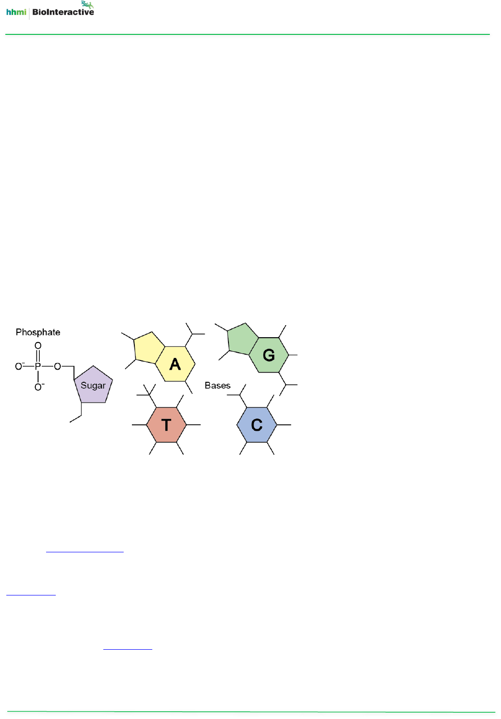
In comparison to proteins, DNA seemed very simple. Frederich Miescher, a Swiss physician, had first isolated DNA
from white blood cells in 1871. Shortly after that, American biochemist Phoebus Levene identified the
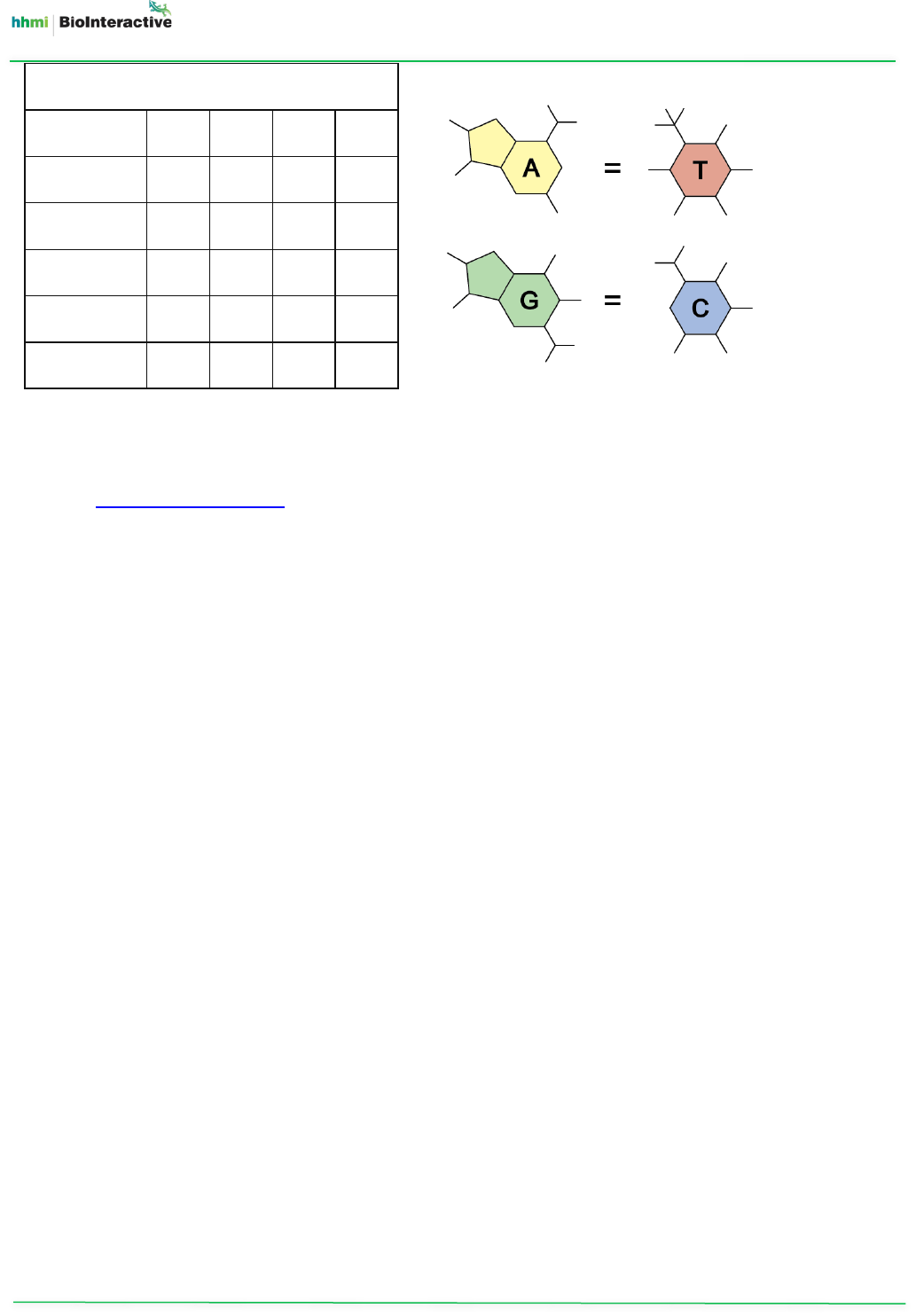
components of DNA: deoxyribose sugar, phosphate, and one of four different nitrogenous bases (Figure 1).
In 1938, British physicist William Astbury took the first x-ray diffraction images of DNA. He used these images to
build a model of the structure of DNA using metal plates and rods. Although his model was very tentative and
contained errors, Astbury correctly positioned the bases lying flat, stacked like a pile of pennies, 0.34 nm apart.
A series of experiments set the stage for establishing that genes were made of DNA and not proteins. Frederick
Griffith’s 1928 experiments showed that pneumococcal bacteria could transfer genetic information between
different strains through a process he called transformation. Oswald Avery, Colin MacLeod, and Maclyn McCarty
determined that the molecule responsible for this transformation was DNA and not protein. Avery and colleagues’
1944 paper was initially met with skepticism, as many scientists continued to believe that proteins were the
genetic material.
In the meantime, more information was emerging about the structure of DNA. American biochemist Erwin
Chargaff reported in a 1950 paper that the proportions of the four nucleotides in a DNA molecule varied among
species. However, within a species, the percentages of adenine (A) and thymine (T) bases were always equal, as
were the percentages of guanine (G) and cytosine (C) (Figure 2). This finding was a key insight for Watson when
he was building his model of DNA.
Figure 1. The nucleotide structure
of DNA. DNA consists of chains of
nucleotides. Each nucleotide is
made of a sugar linked to a
phosphate and one of four bases.
Film Activity
Educator Materials
The Double Helix
DNA & RNA Updated February 2020
www.BioInteractive.org Page 4 of 14
Organism A T G C
Human 30.9 29.4 19.9 19.8
Chicken 28.8 29.2 20.5 21.5
Grasshopper 29.3 29.3 20.5 20.7
Sea urchin 32.8 32.1 17.7 17.3
E. coli 24.7 23.6 26.0 25.7
The most convincing evidence that DNA was the molecule of heredity came from Alfred Hershey and Martha
Chase in a paper published in 1952. Working at Cold Spring Harbor Laboratory in New York, they used radioactive
isotopes of sulfur and potassium to label proteins and DNA, respectively, in bacteriophages. Bacteriophages are
viruses that transfer their genetic material into the bacteria they infect. The Hershey-Chase experiment showed
that the bacteriophage DNA, and not the proteins, entered bacteria for infection.
Modeling DNA’s Structure
At the time of the Hershey-Chase experiment, a number of scientists had started working to determine the
molecular structure of DNA. Among them was Linus Pauling of Caltech, famous for having solved the structure of
several proteins by building models based on chemical bonding principles and biochemical evidence. In 1951, for
example, Pauling had proposed that the polypeptide chains of proteins fold in α-helical structures. Today, the α-
helix is known to form the backbone of tens of thousands of proteins. With his model-building skills, Pauling was
an inspiration to Watson and Crick, as well as the person most likely to solve the structure of DNA before them.
At King’s College London, Maurice Wilkins and Rosalind Franklin were using x-ray crystallography to analyze DNA’s
structure. Despite a few confusing blurry spots, the initial images they obtained hinted that DNA might come in
the form of a twisted spiral, or helix. However, it was not clear how the phosphates, sugars, and bases were
arranged within that helix.
Shortly after Wilkins and Franklin began their experiments, James Watson and Francis Crick decided to work on
DNA as well. Inspired by Pauling’s work, they started building models of DNA molecules. Their approach was to
formulate a possible structure of DNA and then determine whether their model fit experimental observations.
One of their first, and ultimately flawed, models of DNA was a triple-helix model. The triple-helix was made up of
three sugar-phosphate chains. The chains were held together by chemical bonds facilitated by magnesium ions,
and the bases projected outward from this central backbone.
However, Franklin saw that this triple-helix model did not fit the x-ray evidence. Based on her measurements,
DNA fibers contained at least 10 times as much water as Watson and Crick’s model allowed for. Furthermore,
there was no evidence that DNA was associated with magnesium ions. This model also could not explain how the
three phosphate chains could be held together at the center of the molecule.
(Shortly before Watson and Crick produced their successful double-helix model, Pauling produced his own flawed
triple-helix model. Pauling’s model also had the phosphates in the center of the molecule, but without
Figure 2. Examples of Chargaff’s rules. Chargaff discovered that, in a DNA molecule, the proportion of adenine
(A) always equals that of thymine (T), and the proportion of guanine (G) always equals that of cytosine (C).
Relative proportions (%) of bases in DNA

Film Activity
Educator Materials
The Double Helix
DNA & RNA Updated February 2020
www.BioInteractive.org Page 5 of 14
magnesium ions. Instead, Pauling had hydrogen bonds holding the phosphate chains together. Based on what was
known about chemical bonds, however, this model did not work either.)
Franklin’s research provided more insights into the structure of DNA. Her measurements of the water associated
with DNA suggested that the phosphate groups were located in an aqueous environment, likely on the exterior of
the DNA molecule. Improved x-ray images, including Franklin’s famous Photo 51, provided information about the
dimensions of the repeating subunits in a DNA molecule. In addition, her images indicated that DNA molecules
have dyad symmetry, meaning that they look the same when they are turned upside-down or front-to-back
(Figure 3). When Crick found out about DNA’s dyad symmetry, he inferred that the phosphate chains must run in
opposite orientations, or antiparallel, to one another — a brilliant insight that Franklin and others had missed.
Building on these clues and Wilkins and Franklin’s measurements, Watson and Crick once again turned to models
to test their hypotheses of DNA structure. This time, they tried building a double-helix model with two antiparallel
phosphate chains on the outside of the DNA molecule. In this arrangement, the chains would have to be held
together by the bases on the inside, but it was unclear how these bases would pair up.
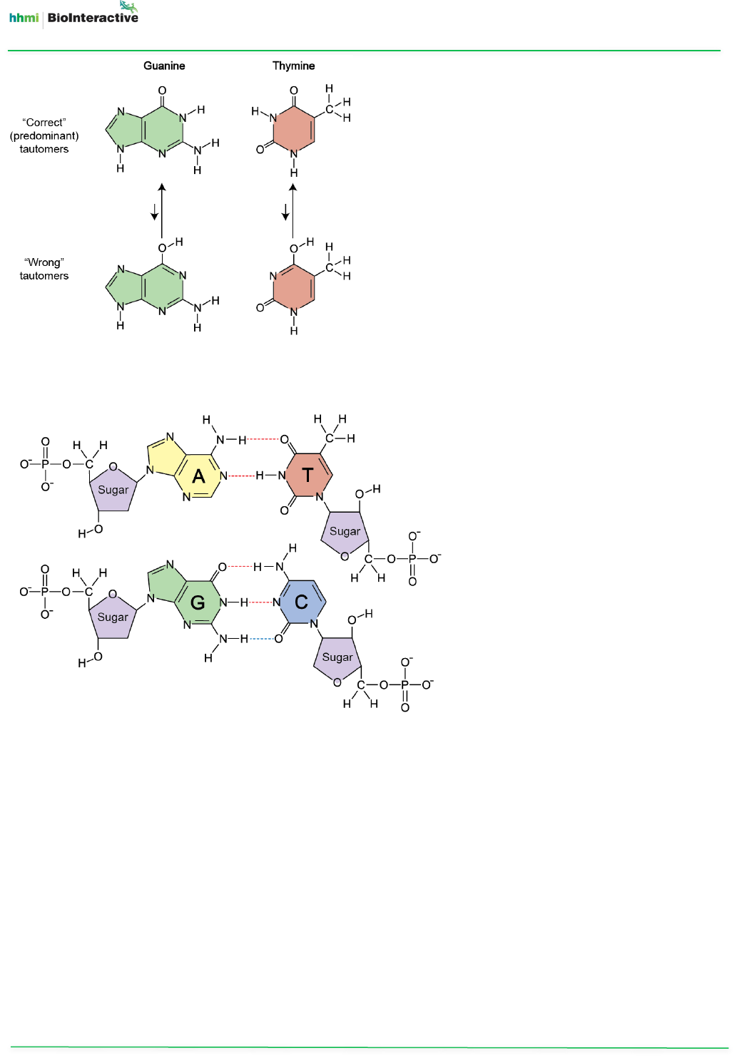
Based on Chargaff’s rules (Figure 2), Crick reasoned that A must always pair with T and G with C. To determine the
bonds between these bases, he and Watson consulted J. N. Davidson’s The Biochemistry of the Nucleic Acids,
published in 1950. However, as with other chemistry textbooks of that time, the book contained drawings of the
“incorrect” forms of guanine and thymine (Figure 4). When Watson and Crick used these forms in their model,
the bases did not form a reasonable hydrogen-bonding pattern (as they would, for example, in the protein
backbone of an α-helix).
A visiting American chemist, Jerry Donohue, helped them correct the structures they were using for the bases,
allowing them to build base pairs with accurate hydrogen bonds. Once Watson incorporated the new, correct
shapes of the bases into his model, he saw where the hydrogen bonds would form, and his and Crick’s model of
DNA quickly fell into place. The A-T and G-C pairings in the new model were consistent with the measurements of
DNA from x-ray images, and the hydrogen bonds between the base pairs made the molecule structurally stable.
C
A
B
Figure 3. This image provides an
example of dyad symmetry.
Whether the original image (A)
is flipped upside-down (B) or
front-to-back (C), it looks the
same.
Film Activity
Educator Materials
The Double Helix
DNA & RNA Updated February 2020
www.BioInteractive.org Page 6 of 14
Watson and Crick initially identified two hydrogen bonds between both A-T and G-C pairings. Today, we know
that there are actually two hydrogen bonds in an A-T pair and three hydrogen bonds in a G-C pair (Figure 5).
The Rise of X-ray Crystallography
Developed early in the 20th century, x-ray crystallography allows the indirect observation of molecular structures
too small to be seen or photographed. The father-son team of William H. and William L. Bragg shared the Nobel
Prize in 1915 for using x-rays to reveal how the repeating structures of crystals form. (William L. Bragg was the
director of the Cavendish Laboratory at the time that Watson and Crick were there. He had been striving to
determine the structures of protein components, but Linus Pauling got there first by discovering the structure of
the α-helix.)
Table salt was the first crystal structure solved by x-ray crystallography in 1914, soon followed by the repeating
carbon structure of diamond. Dorothy Hodgkin and Max Perutz were pioneers in solving the structures of organic
molecules containing more complex atomic arrangements, including cholesterol, penicillin, vitamin B12, insulin,
and hemoglobin.
X-ray crystallography involves mounting a molecule on a stage and bombarding it with a beam of x-rays. The
wavelengths of x-rays are so short that they bounce off atoms within the molecule, “scattering” at specific angles
Figure 4. Different forms, called tautomers, of
guanine and thymine. These bases can have
alternate molecular structures based on different
locations of a particular hydrogen atom. The
tautomers of each base exist in equilibrium, but
one form is more stable and therefore
predominates under the conditions found inside
most cells.
Figure 5. Base pairings in DNA
(hydrogen bonds are shown by
dotted lines). Watson and Crick
rejected the idea of a third
hydrogen bond (bottommost
bond, shown in blue) between
guanine and cytosine because data
hinted that such a bond would be
weak. Later evidence showed that
there are, in fact, three strong
hydrogen bonds in a G-C pair.
Film Activity
Educator Materials
The Double Helix
DNA & RNA Updated February 2020
www.BioInteractive.org Page 7 of 14
that depend on the distances between atoms of various sizes. The scattered x-rays produce diffraction patterns
that can be captured digitally or on photographic film. To interpret these patterns, crystallographers must
determine where x-rays scattered from different atoms overlap. This overlap changes the intensity of spots in the
diffraction pattern.
Two-dimensional images taken at different angles are converted into a three-dimensional model of the molecule
using mathematical calculations called Fourier transformations, which allow the positions of atoms within the
molecule to be determined. When averaged over many observations, these measurements can be accurate to a
fraction of an ångstrom (one 10-billionth of a meter).
The number of x-rays diffracted by a single molecule is too small to observe. Therefore, instead of using a single
molecule, x-ray crystallography examines a sample of many identical molecules. These molecules must be packed
into a highly regular three-dimensional array, so that atoms in all the molecules are arranged the same. If they
aren’t, the x-rays will be bent into overlapping patterns as they travel through the many layers of atoms. This
results in blurry or smeared diffraction patterns that cannot be interpreted or provide very poor resolution.
Crystals have a repeating arrangement of atoms in identical orientations, so they leave a diffraction pattern of
sharp, clear spots. For this reason, biological molecules are typically crystallized, or made to form crystals, before
they are analyzed with x-rays. Scientists may spend much time and effort “growing” crystals of a molecule for x-
ray crystallography, and this can be their rate-limiting step in solving a molecular structure.
Key Evidence for DNA’s Structure Came from X-ray Diffraction
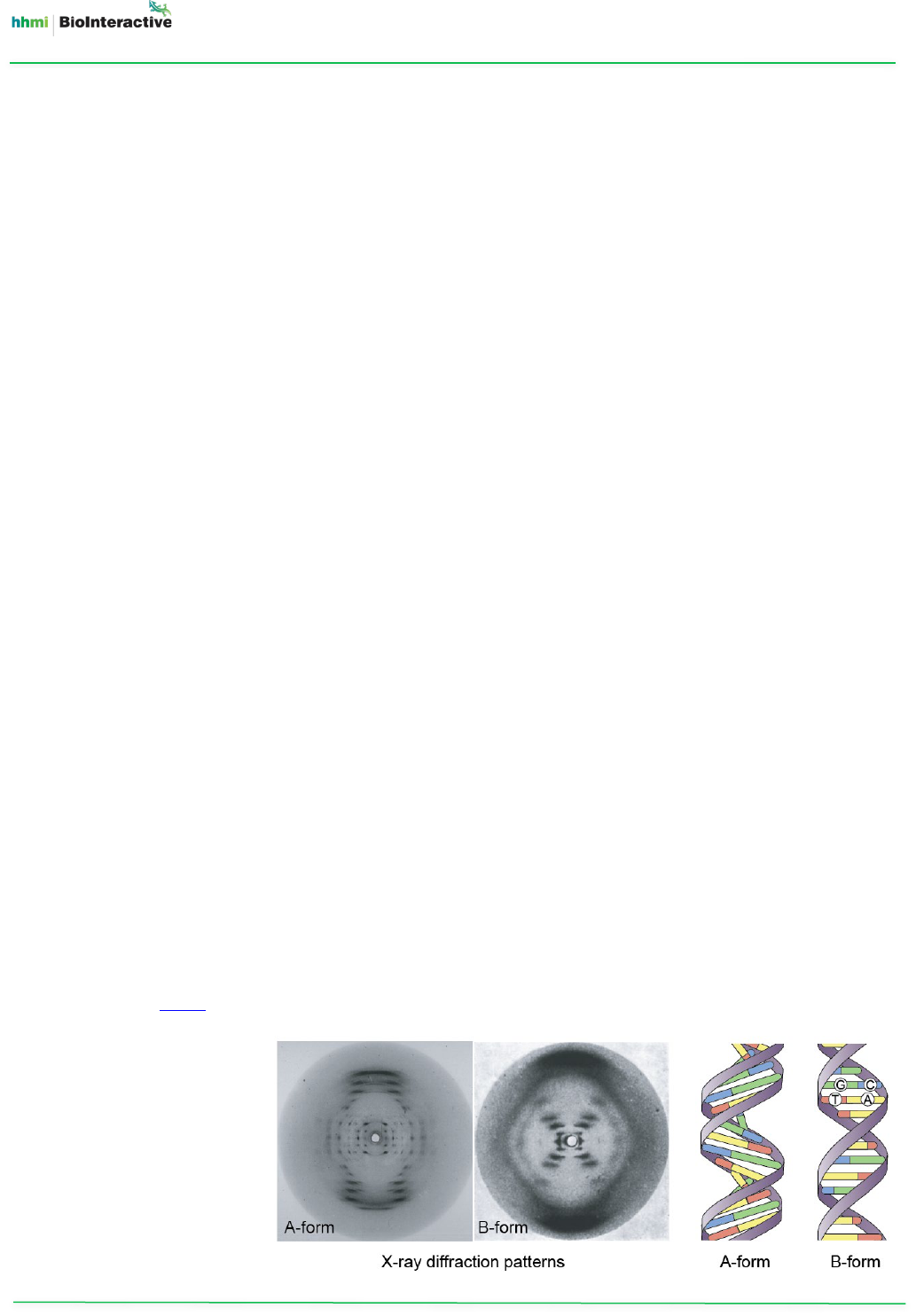
Franklin did not take x-rays of DNA crystals. Instead, she used thin fibers made of thousands of DNA molecules,
which were arranged somewhat like the individual strands of a thick rope, or hair gathered into a ponytail. The
DNA fibers had a sufficiently regular arrangement to produce decipherable diffraction patterns.
By exposing DNA to varying levels of humidity, Franklin and her graduate student Raymond Gosling demonstrated
that DNA existed in two forms, which they called A and B (Figure 6).
• The “dry” A-form occurs when the relative humidity is less than 75%. This form produces a scattered x-ray
diffraction pattern consisting of many distinct spots.
• The “wet” B-form occurs when water molecules cling to the DNA and cause the strands to elongate,
producing an X-shaped diffraction pattern. Because molecules in cells are immersed in liquid, the B-form
is the primary form of DNA inside cells.
(Analyses of DNA crystals, rather than fibers, did not occur until later. It was not until 1980 that an actual crystal
structure of more than a complete turn of B-DNA, in which individual atoms of the DNA could be distinguished,
was published in a paper by Richard Wing and colleagues.)
Figure 6. The A- and B-forms
of DNA. The two forms have
distinct structures, as shown
by their x-ray diffraction
patterns.
Film Activity
Educator Materials
The Double Helix
DNA & RNA Updated February 2020
www.BioInteractive.org Page 8 of 14
Franklin had initially focused her attention on A-form DNA because she thought those images contained more
information. It was, in fact, one of the A-form photos that had revealed that the two strands of DNA ran in
opposite directions — as later realized by Crick.
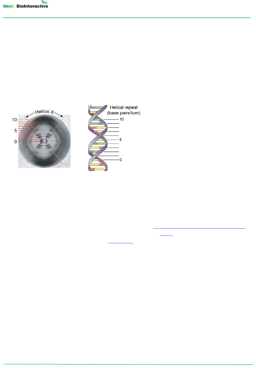
The famous Photo 51 (Figure 7), taken by Franklin and Gosling in May 1952, was of B-form DNA. (As it was the
51st photo taken, Franklin labeled the image 51.) The X-shaped diffraction pattern in the photo is characteristic of
a helical molecule. Independent lines of evidence have confirmed that the diamond shapes formed by the arms
and legs of the “X” indicate the repetition of the helical pattern, as well as the placement of the phosphate sugar
backbone on the exterior of the DNA molecule and the bases in the interior. By analyzing the blurry smears
composing the “X,” scientists have also been able to calculate the dimensions of the DNA molecule: a radius of 1.0
nm, 0.34 nm between base pairs, and 3.4 nm (10 base pairs) within a complete turn of the helix.
Although Watson and Crick based their model on fiber diffraction, their knowledge of the chemical nature of the
components of DNA allowed them to include the positions of atoms and the chemical bonds between them. Their
model was later confirmed by high-resolution x-ray crystallography.
The Discovery of the Structure of DNA Led to Key Insights
On the morning of February 28, 1953, Watson constructed the double helix model of DNA. Two months later,
Watson and Crick published their findings in Nature in a paper titled “A structure for deoxyribose nucleic acid.” In
the same issue, immediately following Watson and Crick’s paper, was a paper written by Wilkins and his
colleagues Alec Stokes and Herbert Wilson. The third paper in the series was written by Franklin and Gosling.
Wilkins’s and Franklin’s papers presented the evidence they had obtained, corroborating Watson and Crick’s
proposed structure of DNA.
Understanding the structure of DNA helped explain how DNA functioned as the hereditary material. Watson and
Crick noted this in their Nature paper: “It has not escaped our notice that the specific pairing we have postulated
immediately suggests a possible copying mechanism for the genetic material.” Because A is always paired with T
and G with C, the order of bases on one strand determines the order on the other. Thus, if a DNA molecule were
unwound, each strand could be copied into a complementary strand, producing an exact replica of the original
molecule. Errors in the copying mechanism could result in mutations, or changes in the DNA sequence, that could
be inherited by future generations.
In 1962, Watson, Crick, and Wilkins were awarded the Nobel Prize in Physiology or Medicine “for their discoveries
concerning the molecular structure of nucleic acids and its significance for information transfer in living material.”
Franklin’s death in 1958 from ovarian cancer precluded her from receiving many of the honors for the discovery
of DNA’s structure, including sharing in the Nobel Prize, which cannot be awarded posthumously.
Figure 7. Photo 51 and the
structure of DNA. The photo
revealed that B-form DNA was
a double helix with 10
nucleotide base pairs within a
complete turn of the helix. The
“X” indicates a helix. The dark
patches indicate the bases.

Film Activity
Educator Materials
The Double Helix
DNA & RNA Updated February 2020
www.BioInteractive.org Page 9 of 14
It took several years of subsequent study, including a classic 1958 experiment by American geneticists Matthew
Meselson and Franklin Stahl, before the exact relationship between DNA structure and replication was
understood.
DISCUSSION POINTS
• The way the story unfolds in the film may give students the impression that each piece of evidence fell into
place one after the other, in a somewhat linear path. In reality, Watson and Crick considered and discussed
most of the pieces simultaneously, as they worked on the mystery of DNA over many months. Their process
was one of trial and error, and repeatedly circling back to make revisions. For example:
o After seeing Photo 51, Watson suspected that DNA might be a double helix. However, he also continued
to consider a triple helix until he built a double-helix model that fit all the known evidence.
o Based on Franklin’s x-ray data, Watson and Crick initially thought that the phosphate chains were on the
outside of the DNA molecule. However, that piece of evidence did not fit until they figured out how the
nitrogenous bases might pair together at the center of the molecule.
• Much has been written about the ethics of how the credit for the discovery of DNA’s structure was shared
among the scientists who contributed to it (for example, the book Rosalind Franklin: The Dark Lady of DNA by
Brenda Maddox). There has also been public controversy about vocal positions that one of the scientists,
James Watson, has taken regarding race, as discussed in this New York Times article
.
o These issues could become part of your discussion with students. It is important to acknowledge that
scientists are people, subject to their personal shortcomings, and accountable for their decisions
regarding ethical conduct and personal prejudices.
• At the beginning of the film, Sean Carroll sets the stage for the unfolding of the story by saying “The three-
dimensional arrangement of atoms in those molecules had to explain the stability of life, so that traits were
passed faithfully from generation to generation, and also the mutability of life.” The notion that heredity
could be explained by the “arrangement of atoms” of molecules was first raised by world-renowned physicist
Erwin Schrödinger.
o In 1944, Schrödinger published the book What Is Life?, in which he argued that living things should be
considered in terms of molecular and atomic structure, as they obey the same laws of chemistry and
physics. According to Schrödinger, genes are passed from generation to generation because the genetic
code was a result of the arrangement of atoms within a molecule. These thoughts inspired a whole
generation of researchers, including Watson and Crick.
o Before your students watch the film, you may want to discuss with them the importance of
understanding three-dimensional molecular structures. Ask them, “How does knowing the structure of
any object tell you about its function?”
• There are many opportunities throughout the film to discuss the nature of scientific inquiry — for example,
how science tries to answer questions about the natural world.
o Lead students in a discussion of the bigger-picture questions that scientists were trying to answer by
determining the structure of DNA. Have them consider that, in the early 1950s, the principles of genetics
were known, but no one knew what the genetic material was (let alone how any physical or chemical
structure was related to the consistency of inheritance, and also the capacity to evolve new traits).
• The film shows how individual scientists take different approaches in trying to understand a process or solve a
problem, due in part to differences in training, the tools available where they work, and their personalities.
Many scientists use a combination of data-gathering and hypothesis-testing approaches. In the film:

Film Activity
Educator Materials
The Double Helix
DNA & RNA Updated February 2020
www.BioInteractive.org Page 10 of 14
o Franklin and Wilkins wanted to solve the structure of DNA by obtaining x-ray diffraction data. They relied
on experiments and observations.
o Watson and Crick built theoretical models which allowed them to see whether those models agreed with
what was known about chemical bonding and x-ray data.
• The film demonstrates how hypotheses must be tested and evaluated against evidence.
o For example, earlier researchers gathered evidence that DNA was the genetic material. Evidence from x-
ray diffraction patterns and Chargaff’s base-pairing ratios supported Watson and Crick’s model of the
structure of DNA.
o Ask students to identify the key pieces of evidence that Watson and Crick used to construct their model
of DNA. Some of the evidence presented in the film includes the structure of the nucleotide (a sugar
linked to a phosphate and one of four nitrogenous bases), Chargaff’s rules (A = T and G = C), and x-ray
diffraction images (showing that DNA is a helix and the molecule’s dimensions).
• The film provides an opportunity to discuss the importance of “failure” in scientific discovery. As biologist
Karolin Luger points out in the film, scientists cannot allow themselves to be paralyzed by the fear of making
mistakes. Although one may want to prove their hypotheses are “correct,” formally speaking, refuting a
hypothesis is also useful. Additionally, as new evidence emerges, models can be modified or sometimes even
rejected.
o Point out to students that Pauling, Watson, and Crick initially hypothesized that DNA was a triple helix.
Although this model was based on sound logic at the time, it ultimately did not fit the data. Watson and
Crick used the data to revise their model.
• The film contains many illustrations and animations of structures of DNA molecules and base pairings. Some
of the DNA animations are based on historical models that have since been refuted. You might use these
animations to have students learn more about molecular structures. For example:
o In the animation in which James Watson is building the DNA molecule and the bases start coming
together, only two hydrogen bonds are shown between bases. Today, we know that there are two
hydrogen bonds in an A-T pair and three hydrogen bonds in a G-C pair (Figure 5).
o In the animation showing the DNA molecule replicating, both strands are being replicated in the same
direction. The mechanism of DNA replication was unknown at the time of the double-helix discovery.
Today, we know that the two DNA strands are replicated in antiparallel directions.
• Students might be interested in learning more about the people featured in the film. For example:
o James Watson is the only scientist involved in the original research who was interviewed in this film. The
interviews with Francis Crick, who passed away in 2004, consist of historical footage obtained when Crick
was at the Salk Institute in California.
o Olivia Judson, the film’s narrator, is an evolutionary biologist based at Imperial College London. She is
well-known for her 2002 book Dr. Tatiana’s Sex Advice to All Creation. In addition, she was featured in the
NOVA documentary What Darwin Never Knew and wrote a weekly blog on evolutionary biology for the
New York Times website.
o Sean B. Carroll, one of the commentators in the film, is a Howard Hughes Medical Institute (HHMI)
investigator and HHMI’s vice president for science education. Carroll is an internationally recognized
evolutionary biologist who studies the way new animal forms have evolved. His research on a wide
variety of animal species has revealed how changes in the genes that control animal development shape
the evolution of body parts and body patterns. Carroll is also well-known for his books Making of the
Fittest, Endless Forms Most Beautiful, Remarkable Creatures, and Into the Jungle. He is also a co-author of
the genetics textbook Introduction to Genetic Analysis.

Film Activity
Educator Materials
The Double Helix
DNA & RNA Updated February 2020
www.BioInteractive.org Page 11 of 14
o Karolin Luger, another commentator in the film, is an HHMI investigator at Colorado State University.
Luger’s research interests include the structural biology of chromatin, the complex of DNA and proteins
that forms chromosomes in the nucleus of eukaryotic cells. In 1997, she determined the structure of the
nucleosome, the basic unit of DNA packaging, which consists of a segment of DNA wrapped around
histone proteins. Using this structure as a starting point, Luger’s work has shed light on how the
nucleosome changes shape, how chromatin interacts with the cell’s transcription machinery, and how
subtle changes in histones can affect overall nucleosome structure.
o Robert Olby, the last commentator in the film, is a science historian at the University of Pittsburgh. He is
the author of The Path to the Double Helix and a biography of Francis Crick entitled Francis Crick: Hunter
of Life’s Secrets.
STUDENT HANDOUT
We designed the student handout as a learning assessment that probes students’ understanding of the key
concepts addressed in the film, which can be used before or during the film to assess students’ prior knowledge
and to guide students as they watch the film. We encourage you to choose the use that best fits your learning
objectives and your students’ needs. Moreover, because the vocabulary and concepts may be complex, we
encourage you to modify the handout as needed (e.g., reducing the number of questions, explanations of
complicated vocabulary for English learner students).
ANSWER KEY
1. In the 1950s, many scientists thought that proteins, not DNA, carried genetic information.
a. Why did proteins seem better suited for storing genetic information?
Proteins seemed better suited because there are many different types of proteins with different shapes and
functions, just like there are many different types of inheritable traits. DNA is simpler and doesn’t vary as
much, so it didn’t seem as useful for storing information.
b. Oswald Avery’s experiments with bacteria led him and other scientists to propose the following claim: DNA,
not proteins, carries genetic information. Complete the table below to explain how Avery’s experiments
supported this claim.
Claim: DNA, not proteins, carries genetic information.
Evidence: (List three pieces of evidence for the claim from Avery’s experiments.)
1.
Avery isolated a substance that transferred a trait from one bacterium to another, which
he called a “transforming principle.”
2.
The transforming principle was not destroyed by a protein-digesting enzyme.
3.
The transforming principle was destroyed by a DNA-digesting enzyme.
Reasoning: (In full sentences, explain how each piece of evidence supports the claim.)
Avery’s experiments showed that DNA, not proteins, carries genetic information. The substance
Avery isolated transferred a trait from one bacterium to another, which showed that the
substance carried genetic information about the trait. A protein-digesting enzyme didn’t
destroy the function of the substance, but a DNA-digesting enzyme did. This evidence showed
that proteins are not important for carrying genetic information about a trait, but DNA is.
2. What are the chemical components of a DNA nucleotide?
a.
a phosphate, a sugar, and a nitrogenous base
b. a phosphate, a nitrogenous base, and an amino acid
c. a nitrogenous base, a sugar, and an amino acid
d. a nitrogenous base, ATP, and a sugar

Film Activity
Educator Materials
The Double Helix
DNA & RNA Updated February 2020
www.BioInteractive.org Page 12 of 14
3. The two strands of a DNA molecule are held together by hydrogen bonds between the:
a. phosphate groups on each strand
b.
nitrogenous bases on each strand
c. bases and the phosphate-sugar backbone
d. carbon atoms in the sugars
4. In the diagram below, Strands I and II represent complementary sections of DNA. The sequence of Strand I is
shown. What is the sequence of Strand II?
Strand I -----------CTAC-----------
Strand II -----------????-------------
a. AGCA b. CTAC c. TCGT d.
GATG
5. The instructions for the traits of an organism are determined by:
a. the proportions of A, T, C, and G in DNA molecules
b.
the order of nucleotides in DNA molecules
c. the length of DNA molecules
d. the way nucleotides are paired in the two strands of a DNA molecule
6. Watson and Crick first built a triple-helix model of DNA. In this model, the nitrogenous bases were on the
outside of the DNA molecule, and the phosphate groups were on the inside.
a. At that time, why did it seem reasonable for the bases to be on the outside of the DNA molecule?
The bases were the most likely parts of DNA to contain genetic information, since they vary in amount
among species and in their arrangement within DNA. So it seemed reasonable that the bases would be on
the outside of the DNA molecule, where they could be accessed more easily by the cell.
b. What evidence caused Watson and Crick to revise this model? Give specific examples from the film.
They found out they had misremembered some of the key measurements from Franklin’s work, so their
original model was based on wrong information. After seeing Franklin’s x-ray images of DNA and her
report on DNA’s symmetry, Watson and Crick built a revised model of DNA’s structure: a double helix with
sugar-phosphate backbones running in opposite directions. They also used Chargaff’s observations of base
ratios to figure out how the bases were paired.
7. Table 1 contains data that Erwin Chargaff published about the composition of DNA.
Table 1. Proportions of nitrogenous bases in the DNA of different organisms. Data from Chargaff and
Davidson (1955).
Organism Tissue % Adenine % Guanine % Cytosine % Thymine
Yeast 31.3 18.7 17.1 32.9
Sea urchin Sperm 32.8 17.7 18.4 32.1
Rat Bone marrow 28.6 21.4 21.5 28.4
Human Thymus 30.9 19.9 19.8 29.4
Human Sperm 30.3 19.5 19.9 30.3
a. Compare the composition of the DNA in the different organisms. Describe any similarities or differences
you observe.
Student answers may vary. They may note that the organisms have different proportions of A, T, C, and G
in their DNA. Within each organism, A and T are present in similar amounts, and the same is true for C and
G. The DNA samples from the two human tissues have almost the same proportions of each nitrogenous
base. The proportions in the yeast and sea urchin samples are also similar to each other.
Film Activity
Educator Materials
The Double Helix
DNA & RNA Updated February 2020
www.BioInteractive.org Page 13 of 14
b. Based on the data in Table 1, mark the following statements as true (T) or false (F). Justify each answer in
one or two sentences.
T
In each organism, there is approximately one adenine for every thymine.
The proportions of A and T are very similar within each organism, so these bases have about a 1:1 ratio.
F
In each organism, the proportions of adenine plus thymine equal those of cytosine plus guanine.
In all of these organisms, A + T > C + G. In the yeast sample, for example, A + T = 64.2% and C + G = 35.8%.
F
In each organism, there is approximately one guanine for every thymine.
The proportions of G and T are not similar within any of the organisms, so these bases do not have a 1:1
ratio.
T
In each organism, there is approximately one guanine for every cytosine.
The proportions of G and C are very similar within each organism, so these bases have about a 1:1 ratio.
c. Why are the proportions of nitrogenous bases in the DNA of two different human tissues (thymus and
sperm) about the same?
All cells and tissues in a particular organism (in this case, a human) share the same DNA sequence.
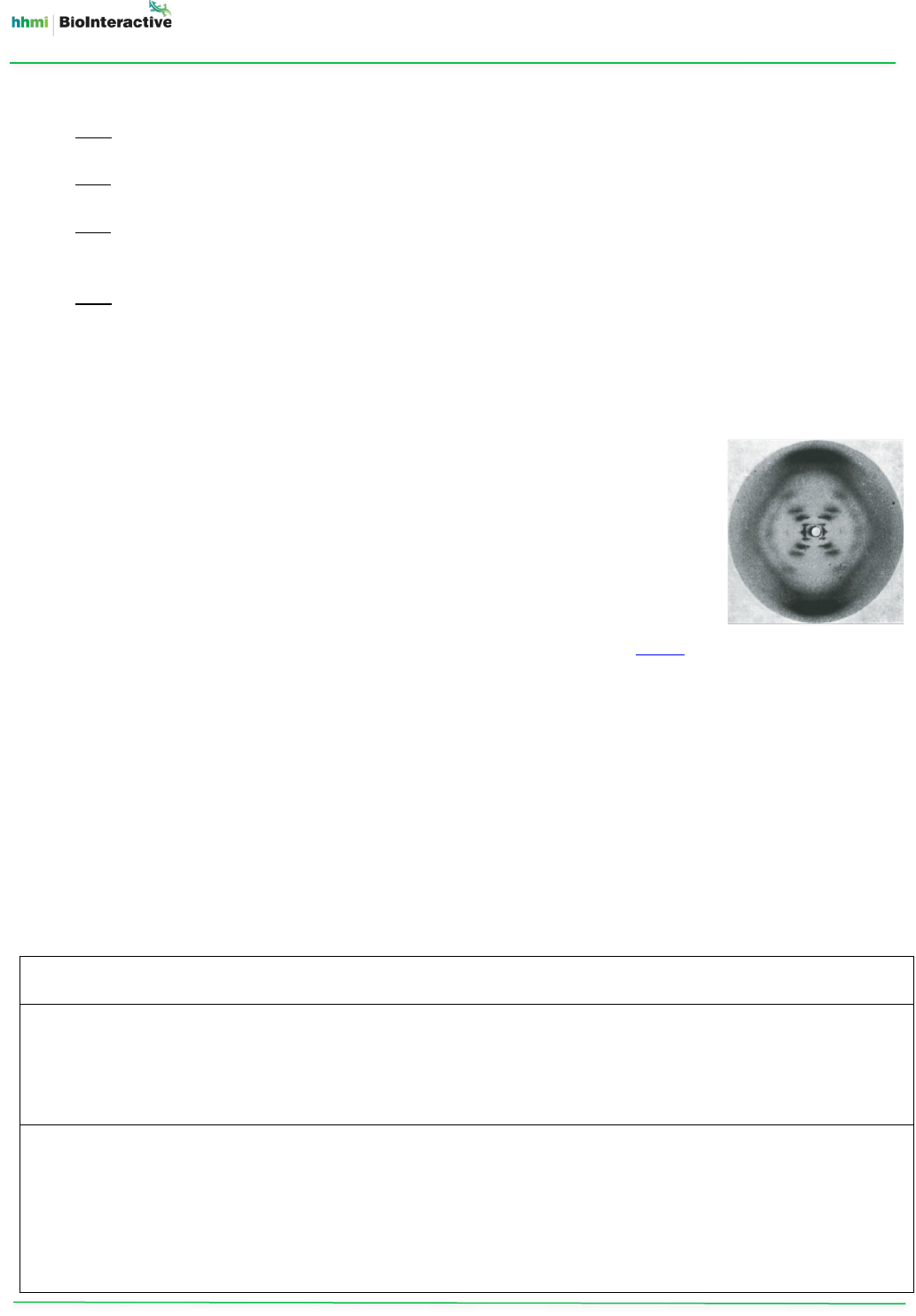
8. The image on the right is of Photo 51, which was taken in 1952 by Rosalind Franklin
and her student Raymond Gosling. It shows the x-ray diffraction pattern of a DNA
molecule, which provides information about the positions of atoms in DNA.
a. Describe the patterns you see in the image.
Student answers may vary. They may note the X-shaped pattern, which has three
or four dark black lines in each arm of the “X.” The angles of the arms above and
below the “X” seem to be similar. The angles of the arms to the right and to the
left of the “X” also seem to be similar. (For more information on how to interpret
this diffraction pattern, consult the “Background” section above and this article.)
b. What conclusions did Watson and Crick reach after seeing this image and reading Franklin’s report
discussing the symmetry of DNA?
Crick had previously worked out that the diffraction pattern of a helix would look like an “X,” so Watson
realized the image showed the diffraction pattern of a helix. Watson concluded that DNA could be a double
helix with two strands. The report from Franklin included an observation about the symmetry of DNA. This
led Crick to conclude that the two strands ran in opposite directions, so the sugar-phosphate backbones
had to be on the outside with the bases inside.
9. Watson and Crick used scientific reasoning, their knowledge of biochemistry, and the research of other
scientists to make one of the most important scientific claims of their time: DNA is a double helix with strands
running in opposite directions. Between these strands, A pairs with T, and C pairs with G. Complete the table
below to explain the evidence that Watson and Crick used to support this claim.
Claim: DNA is a double helix with strands running in opposite directions. Between these strands, A pairs with T,
and C pairs with G.
Evidence: (List three pieces of evidence for the claim. Name the scientists who were responsible for each piece.)
1.
Franklin and Gosling took a photo showing the x-ray diffraction pattern of DNA.
2.
Franklin reported on the symmetry of DNA.
3.
Chargaff found that, in multiple organisms,
DNA had equal amounts of A’s and T’s and equal amounts of
C’s and G’s.
Reasoning: (In full sentences, explain how each piece of evidence supports the claim.)
The research done by Franklin, Gosling, and Chargaff supports the claim that DNA is a double helix with strands
running in opposite directions, between which A pairs with T and C pairs with G. The x-ray diffraction pattern of
DNA, shown by Franklin and Gosling, suggested that DNA was a helix. The symmetry of DNA, reported by
Franklin, suggested that DNA had two strands running in opposite directions, making it a double helix. And
Chargaff’s observation that DNA had equal amounts of A’s and T’s and equal amounts of C’s and G’s suggested
that every A must be paired with a T, and that every C must be paired with a G.

Film Activity
Educator Materials
The Double Helix
DNA & RNA Updated February 2020
w
ww.BioInteractive.org
P
age 14 of 14
10. Even before the structure of DNA was known, studies indicated that the genetic material must have the
following properties:
• be able to store information
• be consistently replicated between generations
• be able to allow for changes, and thus evolution, to occur
Explain how the structure of DNA gives it these three properties. Write one or two sentences per property.
•
The order of the bases (A, T, G, and C) in a DNA molecule stores information.
•
The bases are complementary (A always pairs with T and G with C), so the order of bases on one strand
determines the order on the other strand. So each strand of DNA can be copied into a complementary
strand, producing an exact copy of the original molecule.
•
Errors in the DNA copying mechanism can result in mutations, or changes in the DNA sequence. These
mutations can be inherited by future generations, allowing evolution to occur over time.
REFERENCES
Avery, Oswald T., Colin M. MacLeod, and Maclyn McCarty. “Studies on the chemical nature of the substance inducing
transformation of pneumococcal types.” The Journal of Experimental Medicine 79, 2 (1944): 137–158.
https://doi.org/10.1084/jem.79.2.137.
Chargaff, Erwin. “Chemical specificity of nucleic acids and mechanism of their enzymatic degradation.” Experientia 6, 6
(1950): 201–209. https://doi.org/10.1007/bf02173653.
Chargaff, Erwin, and J. N. Davidson, eds. The Nucleic Acids. New York: Academic Press, 1955.
Franklin, Rosalind E., and Raymond G. Gosling. “Molecular configuration in sodium thymonucleate.” Nature 171, 4356 (1953):
740–741. https://doi.org/10.1038/171740a0.
Griffith, Fred. “The significance of pneumococcal types.” Journal of Hygiene 27, 2 (1928): 113–159.
https://doi.org/10.1017/s0022172400031879.
Hershey, A. D., and Martha Chase. “Independent functions of viral protein and nucleic acid in growth of bacteriophage.”
Journal of General Physiology 36, 1 (1952): 39–56. https://doi.org/10.1085/jgp.36.1.39.
Judson, Horace F. The Eighth Day of Creation: Makers of the Revolution in Biology. Cold Spring Harbor: Cold Spring Harbor
Laboratory Press, 1979.
Meselson, Matthew, and Franklin W. Stahl. “The replication of DNA in Escherichia coli.” Proceedings of the National Academy
of Sciences 44, 7 (1958): 671–682. https://doi.org/10.1073/pnas.44.7.671.
Olby, Robert. The Path to the Double Helix: The Discovery of DNA. Seattle: University of Washington Press, 1974. Revised
1994.
Olby, Robert. Francis Crick: Hunter of Life's Secrets. Cold Spring Harbor: Cold Spring Harbor Laboratory Press, 2009.
Watson, James D. The Double Helix: A Personal Account of the Discovery of the Structure of DNA. New York: Atheneum Press,
1968.
Watson, James D., and Francis H. C. Crick. “A structure for deoxyribose nucleic acid.” Nature 171, 4356 (1953): 737–738.
https://doi.org/10.1038/171737a0.
Wilkins, Maurice H. F., Alexander R. Stokes, and Herbert R. Wilson. “Molecular structure of deoxypentose nucleic acids.”
Nature 171, 4356 (1953): 738-740. https://doi.org/10.1038/171738a0.
Wing, Richard, Horace Drew, Tsunehiro Takano, Chris Broka, Shoji Tanaka, Keiichi Itakura, and Richard E. Dickerson. “Crystal
structure analysis of a complete turn of B-DNA.” Nature 287, 5784 (1980): 755–758. https://doi.org/10.1038/287755a0.
CREDITS
Written by Cindy Gay, Steamboat Springs High School, CO; Laura Bonetta, HHMI; Mary Colvard, Cobleskill-Richmondville High
School (retired), Deposit, New York
Edited by Esther Shyu, Dennis Liu, Eriko Clements, HHMI; Susan Dodge, consultant
Scientific review by Karolin Luger, Colorado State University
Illustrations by Heather McDonald
